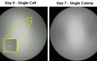
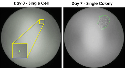
Celigo Provides Image-Based Proof of Single-Cell Clonality
The FDA identifies clonality as one of the most crucial steps in guaranteeing cell line quality and safety. The Celigo Image Cytometer makes single cell sorting easy.
The FDA identifies clonality as one of the most crucial steps in guaranteeing cell line quality and safety. The Celigo Image Cytometer makes single cell sorting easy.
The Danish Cancer Society Research Center recently published a study furthering their analysis of homologous recombination DNA repair machinery. The group previously reported on a growth factor, PSIP1, that enables DNA end resection. With GFP-transfected U2OS cells, the group investigated a structurally similar protein, hepatoma-derived growth factor-related protein 2 (HDGFRP2). The Celigo analyzed cell number and viability via fluorescent markers. The group reports that HDGRFP2 may help to repair silent genes that have been impaired or active genes inhibited by DNA damage. Read the full publication here.
At the Canary Center at Stanford for Early Cancer Detection, investigators studied how AshwaMAX (a steroidal lactone from a winter cherry plant, Withania somnifera, extract) might work as an oral treatment for those with the highly aggressive cancer glioblastoma multiforme (GBM). A heterogeneous disease, non-specific therapies for GBM have proven largely ineffective. Two patient-derived GBM lines (GBM2, GBM39) and one GBM cell line were cultured to create neurospheres that were then exposed to various concentrations of AshwaMAX. Celigo measured cell proliferation and cell death via Trypan Blue staining. AshwaMAX inhibited the neurospheres at nanomolar concentrations. After additional work in vivo, [...]
University of North Carolina researchers investigated different techniques for inhibiting the catalytic activity of protein hTERT – a marker of advanced stage endometrial cancer. Endometrial cancer cell lines ECC-1 and Ishikawa were exposed to either siRNA or a small molecule pharmacological inhibitor BIBR1532, in addition to the drug paclitaxel, to see whether inhibiting hTERT provided additional efficacy against these cancer cells. The Cellometer, in combination with propidium iodide and Annexin-V FITC, calculated apoptosis in the various treatment conditions. The hTERT inhibition plus paclitaxel did prove synergistic, reducing cell growth and invasion more than paclitaxel alone. Furthermore, BIBR1532 antagonized cell invasion [...]
Emily Whitehead, cancer survivor, holding Nexcelom cell counting sheep. Thanks to the lifesaving T cell therapy clinical trial at Children’s Hospital of Philadelphia, Emily Whitehead is now two years cancer free. Diagnosed with acute lymphoblastic leukemia (ALL) right after her 5th birthday, doctors discovered that Emily’s leukemia was particularly resistant to chemotherapy, as are roughly 15% of the total number of ALL cases. After two recurrences of the disease, Emily’s parents enrolled her in a clinical trial for CTL019, an experimental therapeutic using a patient’s own reprogrammed T cells to eliminate the cancer cells. Now 9 years old, [...]
The MD Anderson Cancer Center worked in collaboration with Nexcelom to create a new method by which to measure the cytotoxic potential of natural killer (NK) cells. The traditional non-radioactive method, calcein release, is subject to variation, with differing dynamic ranges depending on tumor type. The Cellometer Vision and calcein, in combination with K562, 721.221, and Jurkat cells, were used to develop a novel, image cytometry-based assay to ascertain NK cytotoxicity. Using fluorescent intensity gating to ignore dimmer cells and apoptotic bodies, image cytometry provided a way to measure tumor cell lysis in a specific manner with the same experimental [...]
Join us for our next Cellometer User Informational Webinar on Friday October 24th at 11am: The Importance of Proper Focus on Cellometer Cell Counters This informational webinar will cover: -a brief introduction to image cytometry -why a good image is needed -why proper focus is so important -time for questions and answers Register here: https://www3.gotomeeting.com/register/343244614 All webinars are recorded. If you know you cannot attend but are interested in the topic, please register. After the webinar is concluded, you will receive an email with a link to view the recorded presentation.
Patients with cancers such as multiple myeloma, leukemia, lymphoma, and other metastatic cancers have tumor cells with unique immunological targets that when exploited, can lead to the complete destruction of the mass and a lasting remission from disease. Clinicians attack those targets using cell therapy, also known as “targeted immunotherapy”. Cell therapy is the programming of a patient’s own immune cells to target that patient’s tumor cells for destruction. One application of cell therapy, Adoptive Cell Transfer (ACT), employs select methods of genetic manipulation and propagation of those immune cells ex vivo so that the newly programmed cells can be [...]
Cellometer Vision CBA combines the simplicity of image cytometry with the power of flow analysis software to offer simple, accurate cell-based assays. Generate comparable results to flow cytometry with the Vision CBA Analysis System: Apoptosis Cellometer Vision CBA video demonstrations Autophagy Cell Cycle Proliferation Transfection Viability ... and Others Features of the Vision CBA Analysis System Dual-Fluorescence: The Vision CBA Analysis System comes equipped with two standard fluorescent optics modules for dual-staining analysis of primary cells in heterogeneous samples. Fast Results: Obtain cell images, counts, size measurements, viability calculations, and population data in < 3 minutes. Enhanced Sensitivity: [...]
This presentation will focus on using Cellometer image cytometry to effectively determine GFP or other fluorescent protein transduction/transfection efficiency and cell concentration, as well as detect and quantify dual expression in a cell population. Quantifying GFP can be done in four easy steps. First pipette 20 ul of target cell sample into a disposable counting chamber. Then insert the slide into the instrument, select your assay from the drop-down menu, focus and click count. Within seconds the instrument acquires images, identifies cells with and without fluorescence and automatically tabulates the results. The results include the number of cells counted in [...]